・韓国・ソウル市・延世(ヨンセ)大学歯科病院 訪問記
2026年5月6日、韓国・ソウルのヨンセ大学を訪問する光栄に浴しました。
この訪問は、愛知学院大学の村上弘教授の退職記念祝賀会にて、数年ぶりにAdvanced General Dentistry科のキム教授と再会したことがきっかけで実現いたしました。
近年、ヨンセ大学歯学部は、QS世界大学ランキング(歯学部門)において、韓国国内で常にトップクラスにランクインするなど、世界的に高い評価を得ています。
これは同大学の傑出した国際的評判と学術的卓越性を反映したものです。
今回の訪問で、キム教授が統合診療科を創設されたことを伺いました。
3室の専用手術室と多数の高度な治療ユニットを備えた臨床施設の規模と洗練さには、ただただ驚かされるばかりでした。
キム教授にご挨拶した後、同科のヤン教授に病院内を親切にご案内いただきました。
ヤン教授がかつて愛知学院大学に留学されていたこと、そして両大学が現在も姉妹校の関係にあることを知り、大変嬉しく思いました。
こうした学術的な絆が、今回の有意義な訪問の礎となったことを、非常に幸運に感じております。
キム教授、そしてヤン教授、皆様の温かいご厚情に改めて感謝申し上げます。
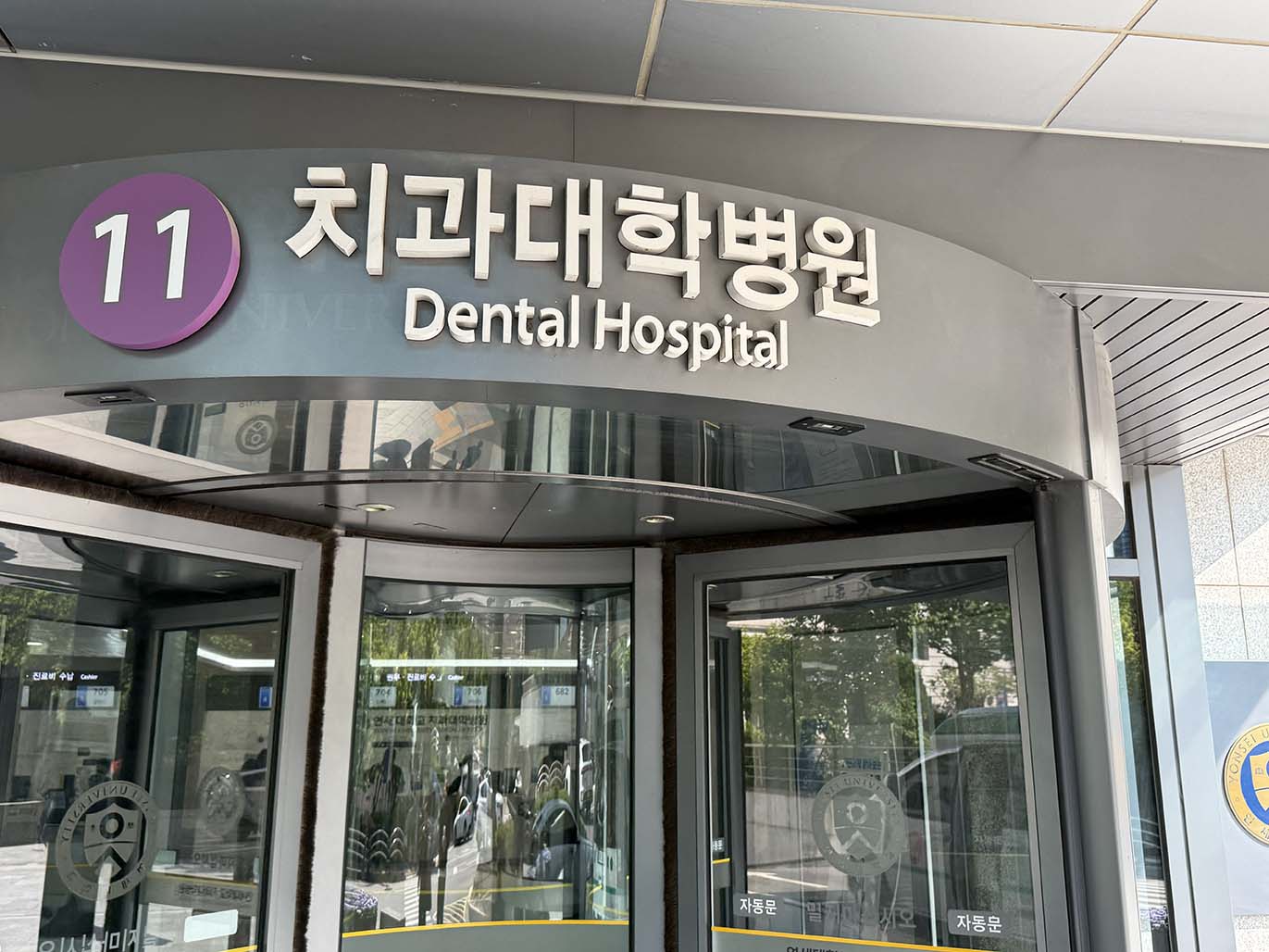
歯科病院玄関です。
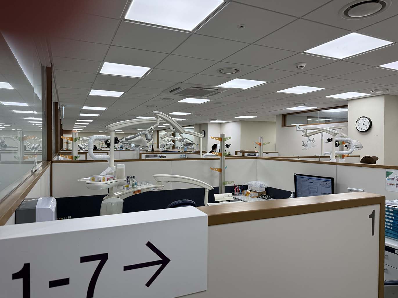
1階ののAdvanced General Dentistryの診療室。
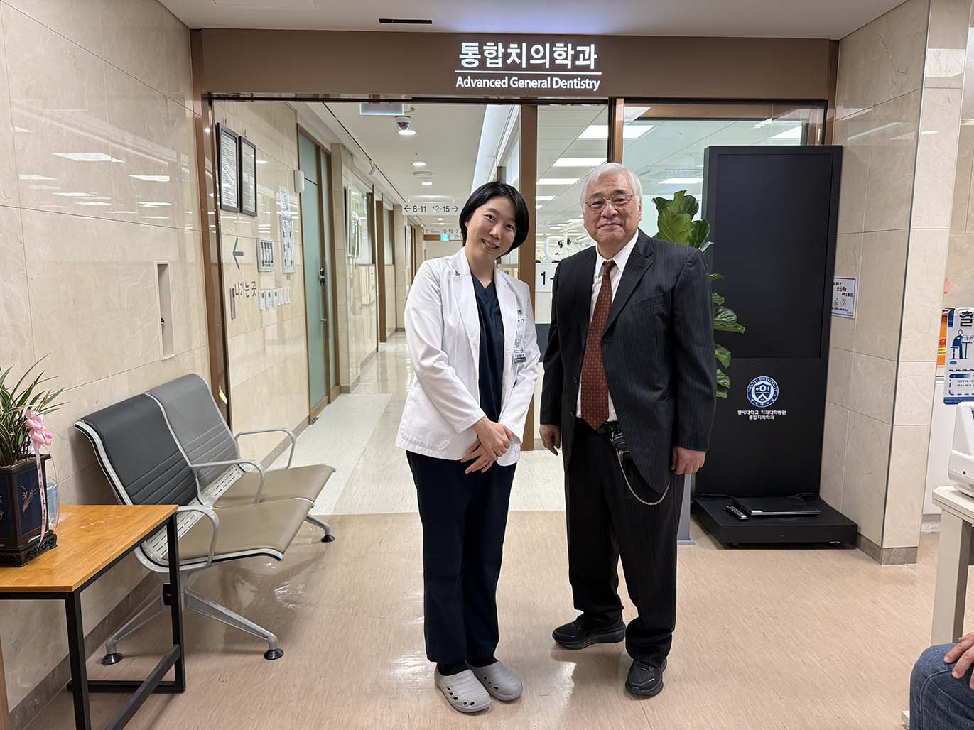
病院内を案内していただいたProf. Yang先生との記念写真。
お忙しいところ、本当にありがとうございました。
ご迷惑をおかけしたと思います。
・27年ぶりに、母校である愛知学院大学歯学部の歯学会誌に共同で症例報告を投稿し、掲載されました。
堀田久斗,堀田康記;上顎前歯部GBRで広範な顔面腫脹および眼瞼部皮下出血斑を生じた1症例:愛知学院大学歯学会誌,63(3):152~158,2025
Hotta H, Hotta Y: A Case of Extensive Facial Swelling and Periorbital Ecchymosis
Following Guided Bone Regeneration in the Anterior Maxilla; The Aichi gakuin
Journal of Dental Science, Vol.63(3), 152-158, 2025, Japan.
In cases of severe anterior maxillary bone resorption, guided bone regeneration
(GBR) is commonly used to achieve functional and esthetic outcomes. Postoperative
swelling is usually mild and self-limiting; however, extensive facial swelling
with periorbital ecchymosis is extremely rare.
We report a 54-year-old non-smoking female with severe periodontal disease
affecting teeth 11–22. Tooth 12 had been previously extracted. Her medical
history included obsessive–compulsive disorder treated with long-term bromazepam.
After extraction of 11–22, GBR was performed using carbonate apatite
(Cytrans®) mixed with platelet-rich fibrin (PRF) and covered with PRF membranes.
Primary closure was achieved with periosteal-releasing incisions, and intraoperative
bleeding was minimal. The following day, the patient developed diffuse
facial swelling and extensive ecchymosis extending to both upper and lower
eyelids. No signs of arterial bleeding, infection, or visual disturbance
were noted. Swelling improved by day 2, and facial appearance was largely
normal by day 9. Blood tests showed mild inflammatory changes without coagulation
abnormalities.
At 5 months, CBCT confirmed sufficient bone regeneration, and four implants
(Straumann Bone Level Tapered, Ø3.3 × 10 mm) were placed uneventfully.
The extensive ecchymosis was likely multifactorial, involving benzodiazepine-related
muscle relaxation, NSAID-induced platelet dysfunction, psychological stress,
and anatomical spread through the SMAS.
Despite its dramatic presentation, the hemorrhage was likely venous
and self-limiting. This case highlights the need for careful preoperative
assessment and awareness of rare but significant postoperative swelling,
particularly in patients with complex medical backgrounds.
・ハノイ医科大学歯学部訪問記
2026年8月9日・10日の2日間、ベトナムのハノイを訪問しました。
9日は、まずハノイ医科大学から徒歩圏内にあるHerident Dental Clinicを訪れ、院長のDr. Đặng Triệu Hùng先生による骨移植手術を見学させていただきました。
同院は7階建てのビルで、約50名のスタッフが勤務しているとのことでした。歯科医院というより、もはや病院と呼ぶべき規模です。
Hung先生は、インプラントロジストというよりも、口腔顎顔面外科専門医と紹介する方が正確でしょう。ハノイ医科大学では口腔病理・顎顔面外科学講座の主任を務め、同大学附属病院では歯科部門の副部長を務めておられます。日常的に手がけているのは、腫瘍切除や顎骨再建など、一般の開業医の診療範囲をはるかに超える症例です。
講演会では、最初に私が90分間の講演を行い、続いてHung先生が腫瘍および顎骨再建について講演されました。先生はこれまでに、このような手術を数千症例経験されているとのことでした。口腔顎顔面外科の高度な内容に触れることができ、大変勉強になりました。
翌日の10日は、ハノイ医科大学歯学部を見学しました。歯学部はハノイ医科大学を構成する一部門です。建物は4階建てですが、各階のフロア面積は広く、最新の設備が整えられていました。夏休み中のため学生の姿はありませんでしたが、充実した教育・診療環境を実際に確認することができました。
ハノイ医科大学は、ベトナム国内でも屈指の難関大学であり、その歯学部も最難関の一つです。歯学部の建物全体を案内していただいた後、隣接する大学附属病院も見学しました。
そこでHung先生と再会し、歯科病棟も案内していただきました。病床は満床でした。
まさに「医療現場の最前線を見た」という印象でした。
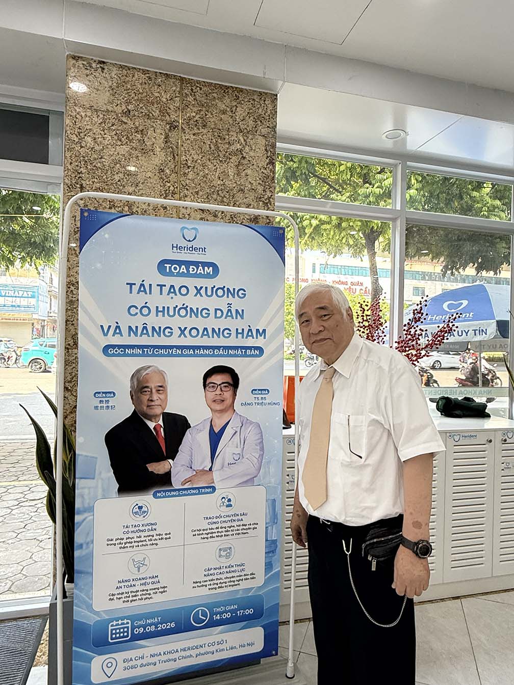
Herident Dental Clinicの玄関にいきなり自分の写真があり、びっくりしました。
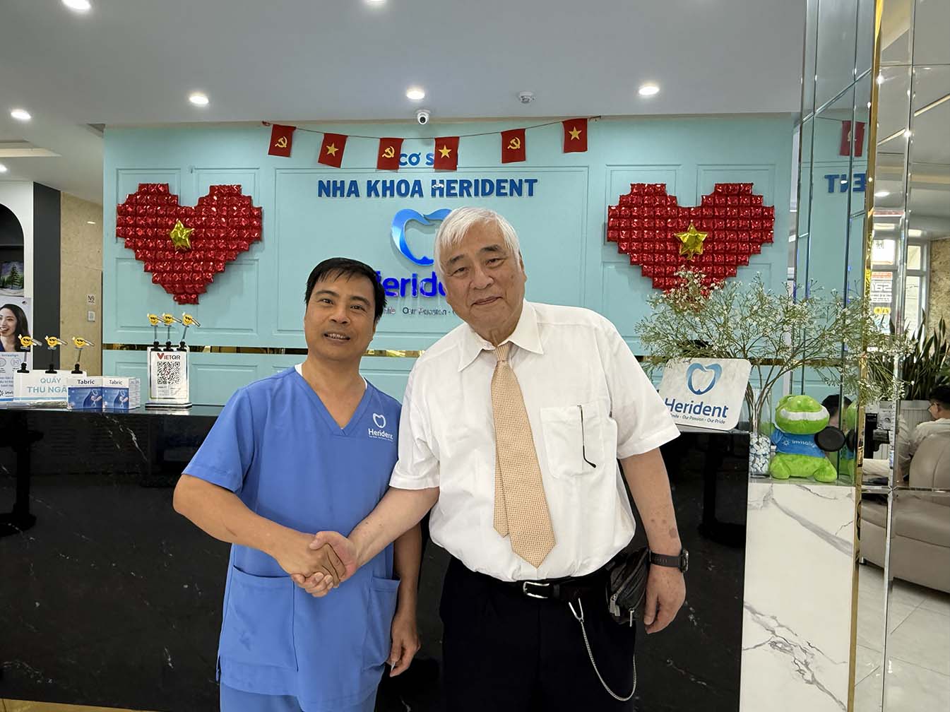
受け付けで、院長のĐặng Triệu Hùng先生とまず挨拶をさせていただきました。
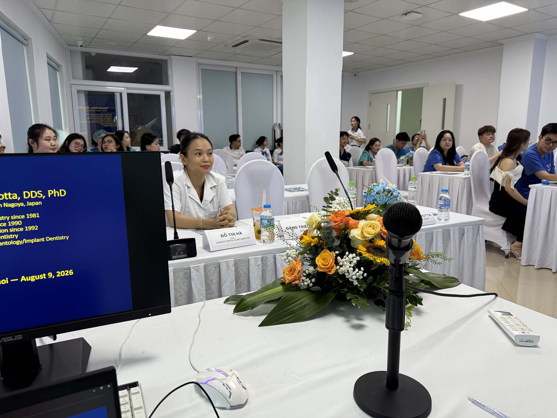
ビル内の講演会場です。もはや大学のクラスル-ムという感じです。
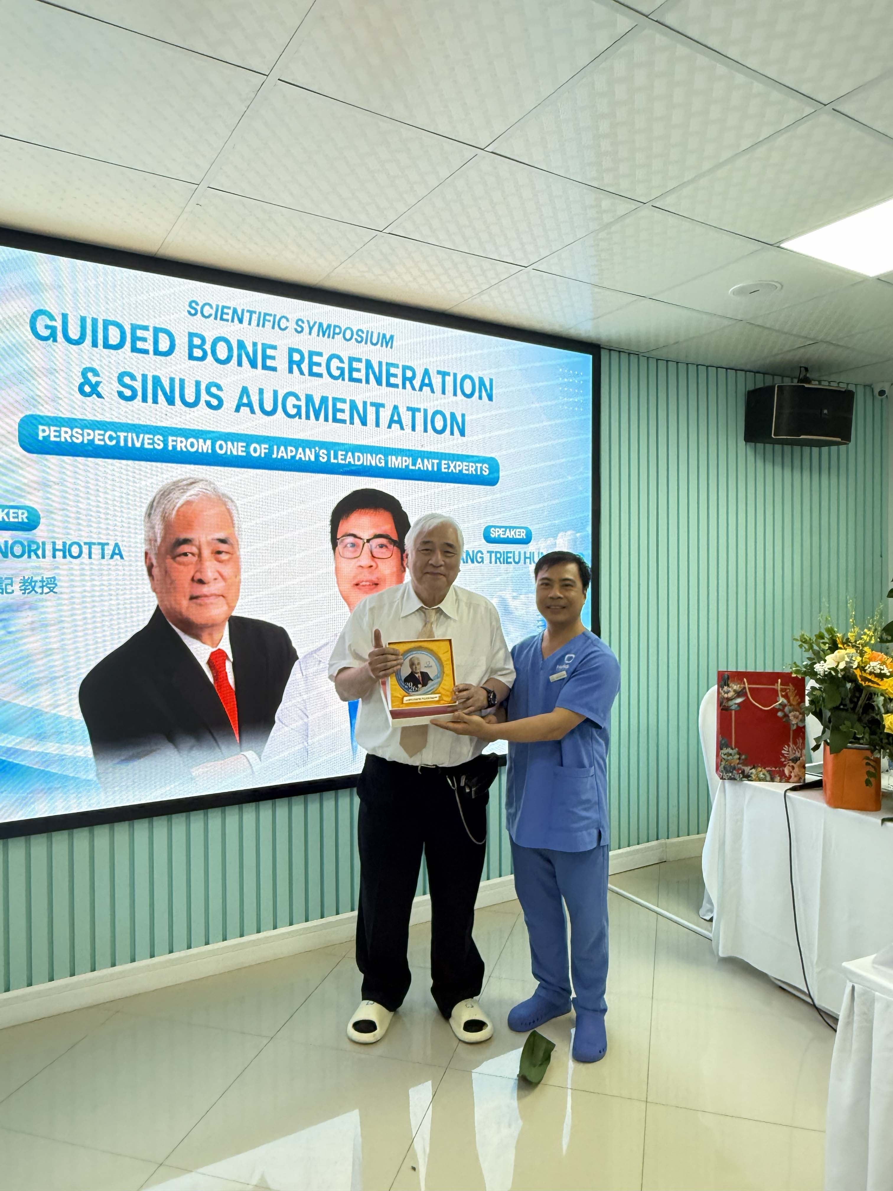
講演後、Đặng Triệu Hùng先生から、クリスタル製の記念盾を頂きました。
恐縮です。ありがとうございます。
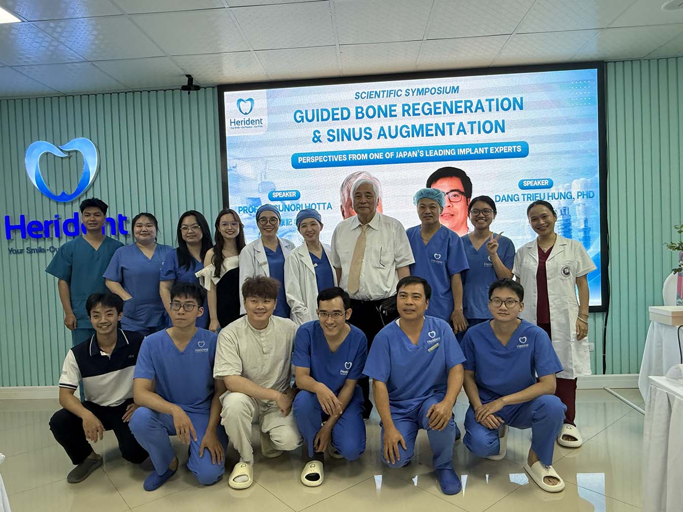
講演後、主なスタッフとの記念写真です。
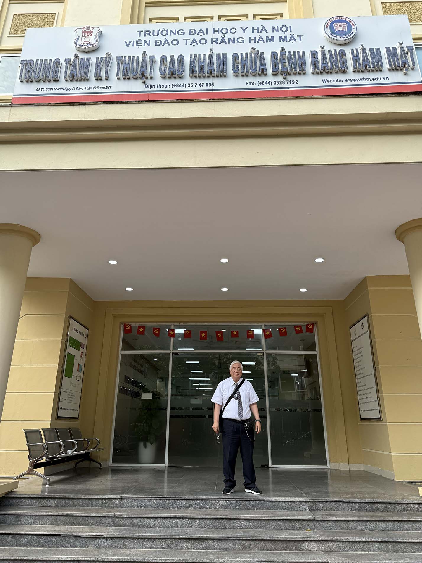
ハノイ医科大学歯学部の玄関です。
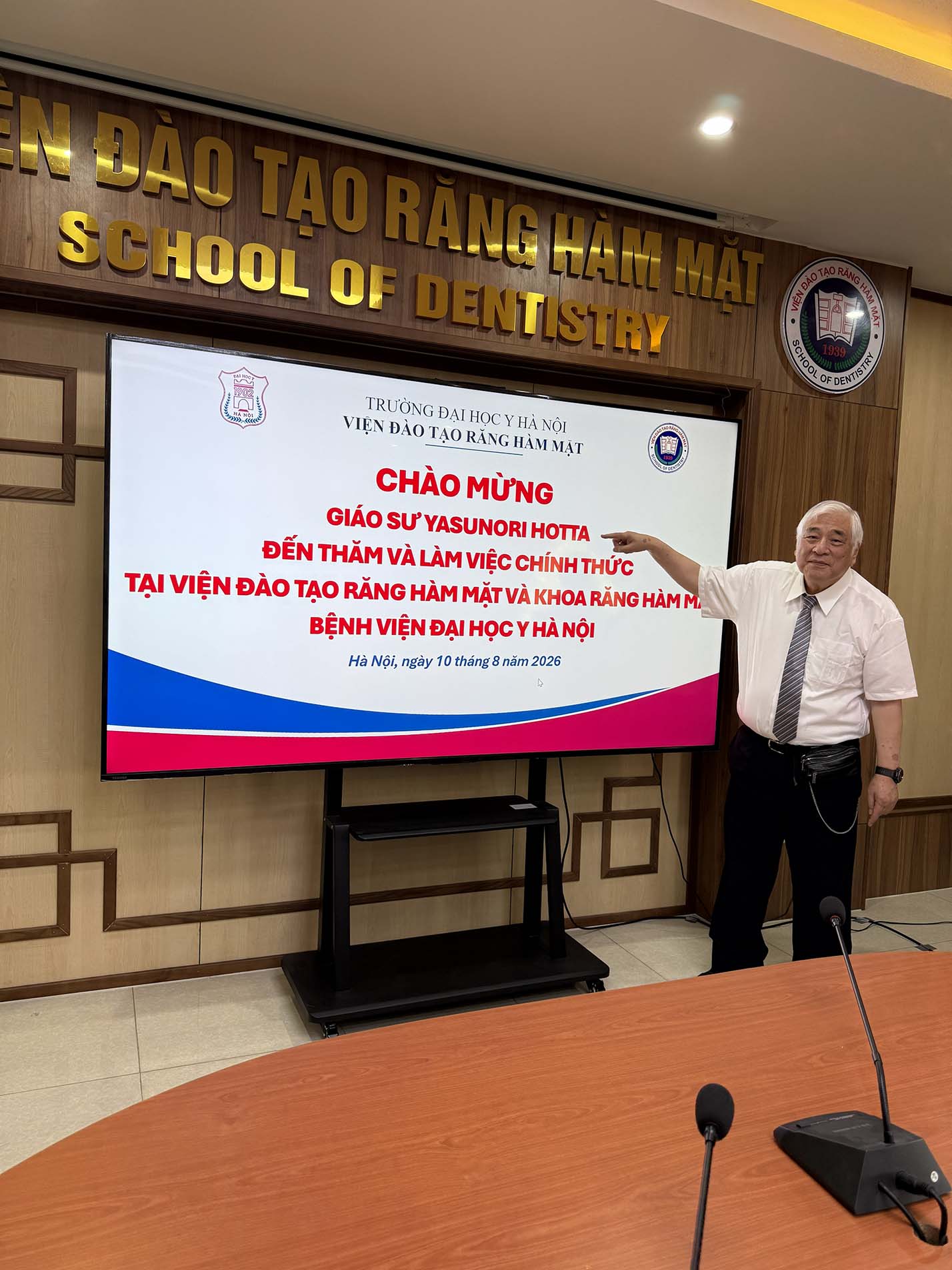
まず応接室に案内されたところ、正面の大型モニターには、
私の名前を記した歓迎メッセージが映し出されていました。光栄です。
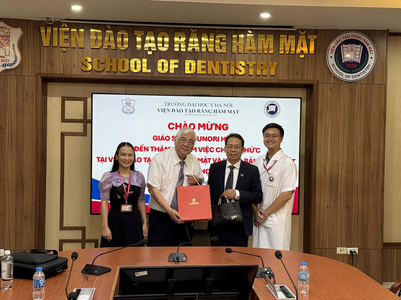
向かって右側(私の左手側)が歯学部長のTong Minh Son先生、反対側の女性がBinh Pham先生です。ハノイ医科大学は、私の母校である愛知学院大学と1992年から姉妹校提携を結び、長年にわたり教育・学術交流を続けています。愛知学院大学歯学部への留学経験を持つBinh先生には、今回のハノイ訪問をコーディネートしていただき、多大なるご尽力を賜りました。皆様には、心より感謝申し上げます。
・2026年5月に実施されたAAID(アメリカ口腔インプラント学会)認定試験のOral Examinationの結果が発表されました。
Associate Fellow受験者2名、Fellow受験者1名の計3名が、全員合格されました。
Nagoya Japan MaxiCourseのDirectorとして、安堵するとともに大変うれしく思います。
認定証は、10月に米国ナッシュビルで開催されるAAID第75回年次学術大会において授与されます。
10月3日午後のBusiness Meetingでは、合格者一人ひとりに認定証が手渡される晴れの舞台が用意されています。
皆様のご努力に心より敬意を表し、改めてお祝い申し上げます。
・JOI(AAID;アメリカ口腔インプラント学会誌)に、論文(Clinical Case Report)が掲載されました。(近々、open accessになります。)
・Hotta H, Hotta Y, Ito K, Komatsu S, Saito T, Teramoto Y. Implant Treatment
for Severe 3-Dimensional Bone Defects in the Area of Missing Adjacent Maxillary
Central and Lateral Incisors: Long-Term Follow-Up Using Titanium Mesh and
GBR Techniques. J Oral Implantol. 2026 Aug 3;52(4):251-260.
Abstract
This report presents 2 cases of severe bone defects following the extraction
of adjacent maxillary central and lateral incisors. In both cases, the
teeth were extracted in the hospital's oral surgery department due to extensive
apical periodontitis. Postoperatively, 3-dimensional bone defects were
observed, including mesiodistal, labial, and palatal bone loss extending
near the nasal floor.
In the first case, 2 implants were placed using the guided bone regeneration
(GBR) method with titanium mesh. Subsequently, during the second stage
surgery, a minor GBR procedure was performed to improve the contour of
the implant neck.
In the second case, in which the bone defect was even more severe than
in the first, a multi-GBR approach was selected. After the initial GBR
procedure, insufficient bone formation was observed in the central area
of the defect. To address this, the ice cream cone technique was employed
to enhance bone regeneration. Subsequently, 2 implants were placed. After
implant placement, a vertical concavity was observed between the necks
of the 2 implants. To correct this, a second GBR procedure using titanium
mesh was performed as in case 1. However, an interimplant black triangle
was observed, and a vascularized interpositional periosteal-connective
tissue flap was performed to eliminate it.
Both cases have been followed for more than 6 years since the placement
of the final prostheses with no significant findings in intraoral examinations
or cone beam computerized tomography images. Esthetically, no interimplant
black triangle was observed.
In case 1, a satisfactory outcome was achieved by remaking the prostheses.
However, in case 2, which had a more severe bone defect than case 1, the
clinical crown length of the prostheses is slightly longer compared with
the contralateral side.
・私のインプラント症例集がクイントより出版されますので、ご案内します。
このたび、クインテッセンス出版より、私の著書『インプラント治療の歴史と実績-43年間の症例写真が語る-』が出版されることとなりましたので、ご案内申し上げます。
本書は、私にとって人生で初めて出版する単行本です。2024年2月に執筆を始め、出版まで約2年半を費やして、ようやく完成させることができました。
私はこれまで、日本および米国の学術誌に、主として症例報告(Case Report)を発表してきました。しかし、一つの論文で伝えられる内容には限りがあります。本書は、それらの症例も含め、私が1981年にインプラント治療を始めて以来、44年以上にわたって経験してきた臨床を、約200ページにまとめたものです。
私にとって本書は、これまでの臨床人生の集大成であると同時に、「臨床家としての遺書」という性格も持つ一冊です。
骨造成、審美領域、重度骨欠損症例などを中心に、長期間にわたって経過を観察してきた症例を収載しています。
また、愛知インプラントインスティチュートの「100時間コース(認定講習会)」において、長年にわたり講義してきた内容をまとめたものでもあります。そのため、卒業生の先生方には、講習会で一度はご覧になったことのある症例も含まれていると思います。懐かしく思い出していただける症例もあるかもしれません。
【本書の目次】
第1章 ブレードインプラント
第2章 インプラントオーバーデンチャー
第3章 歯内骨内インプラント
第4章 骨膜下インプラント
第5章 スプリットクレスト
第6章 GBR
第7章 上顎洞底挙上術
第8章 多数歯欠損症例
第9章 矯正とインプラント治療
第10章 インプラント周囲炎とリカバリー
第11章 抜歯即時埋入と即時荷重
本書は文章中心の専門書ではなく、そのほとんどが実際の症例写真で構成されています。写真を見ながら治療の経過を追うことができますので、専門書ではありますが、比較的気軽に読み進めていただける内容になっていると思います。
単に成功症例を集めたものではありません。治療に悩み、迷い、ときには反省しながら、患者さんとともに歩んできた臨床の記録です。長期経過を通して、診断や治療計画をどのように考えたのか、その思考過程もできる限り率直に記載しました。
本書の執筆にあたっては、長年にわたり愛知インプラントインスティチュートを支えてくださった会員の皆様との出会いや交流が、大きな支えとなりました。改めまして、皆様に深く感謝申し上げます。
本書の発行は、限定500冊のみとなります。利益を得ることを目的とした出版ではありませんので、一般の書店やオンライン書店などの販売ルートには載せず、私から直接お申し込みいただいた先生方にのみ発送いたします。
もとより、500冊すべてが売れるとは考えておりません。売れ残りによる赤字も覚悟したうえで、自分が歩んできた臨床の軌跡を一冊の記録として残したいとの思いから出版いたしました。
もし内容にご関心をお持ちいただけましたら、お手に取っていただければ大変うれしく存じます。もちろん、ご購入を無理にお願いするものではございません。
なお、本書に関する私の著者印税はゼロであり、販売による利益も一切ありません。書籍代、送料等の実費のみでお渡しいたします。
【お支払い金額(1冊)】
書籍原価 13,200円
消費税 1,320円
レターパック送料 430円
事務手数料 50円
────────────────
合計 15,000円
ご購入を希望される先生は、まず、このメールへの返信にて、以下の内容をお知らせください。
・先生のお名前
・必要冊数
・送付先のご住所
・サインの要否
メ-ル確認後、振込先銀行をお知らせします。
本が私の手元に届くのは、9月30日の予定です。ご入金を確認のうえ、準備が整い次第、順次発送させていただきます。
また、ご迷惑でなければ、先生のお名前と私のサインを本書に入れて発送させていただきます。
サインが不要の場合は、その旨をお知らせください。
本書が、先生方の日々の診断や治療計画を考える際の一助となり、インプラント治療の本質について改めて考えていただく機会となれば、著者としてこれ以上の喜びはありません。
愛知インプラントインスティチュート
施設長 堀田 康記
注文のための E-mail: info@hotta-dc.com